Head trauma and concussions caused by contact sports are a quickly growing epidemic among young athletes. When left undetected, concussions can result in long-term brain damage and may even prove fatal. The Center for Disease Control reports show that the number of reported concussions has doubled in the last 10 years. The American Academy of Pediatrics has reported that Emergency Room visits for concussions in kids ages 8 to 13 years old has doubled, and concussions have risen 200 percent among teens ages 14 to 19 in the last decade.
While the first hit can prove problematic, the second or third head impact can cause permanent long-term brain damage. Cumulative sports concussions have been shown to increase the likelihood of catastrophic head injury leading to permanent neurologic disability by 39 percent.
Once a diagnosis of concussion is established, traditional concussion treatment focuses primarily on observation for the first 24 to 48 hours, medication for pain, ice packs or application of a damp cloth to reduce swelling, monitoring for any extreme changes in body temperature, and plenty of rest from mental and physical activities. Although these recommendations are non-invasive and possibly have some palliative benefit, they do not address the underlying core issues.
An innovative medical approach that integrates specialty technology involving osteopathic and chiropractic cranial manipulation coupled with nutritional support provides solutions and resolutions for the underlying causes of concussion. Applying these innovative technologies has resulted in the quick recovery of many chronic concussion patients from their symptoms even after 20 years of suffering post-concussion sequelae.
This newer technology views concussion from two perspectives. First, physiologically and second structurally. Physiologically the brain undergoes physical trauma which can result in bleeding, swelling and neurological damage to the brain cells due to a build up of toxic metabolic waste products. Second and unknown to most medical practitioners there is a major disturbance in the basic motion of the base of the skull with breathing. This latter aspect has been totally ignored because most practitioners are not aware that this process even exists. This discovery was shared with me in 1983 while attending a post-graduate chiropractic seminar. Simplistically, the base of the skull (formed by the junction of the occipital and sphenoid bones) moves into flexion upon inhalation and moves into extension with exhalation. When a concussion occurs, this process gets reversed and places excess tension on the entire dural tube, which attaches around the brain, exits through the foramen magnum, attaches tenaciously to the upper three cervical vertebrae and extends all the way down to the second sacral tubercle. A trauma-induced reversal of this motion is the underlying cause of the common symptoms associated with concussion:
- Headache: Excess tension on the intracranial dural membranes elicits headache because the dura is innervated with sensory nerves from the three branches of the fifth cranial nerve, (trigeminal nerve) and cervial nerves 2 and 3 in the upper cervical area.
- Decreased Cognitive Function: Disruption of the basic motion of the skull disturbs the cerebrospinal fluid flow around the brain. The induced trauma also creates intracranial swelling. This also contributes to brain and cognitive dysfunction by decreasing the level of oxygen, of nutrients available to brain cells, and increase in the accumulation of metabolic toxins. The latter can affect the neuronal pathways causing all types of bodily dysfunctions.
- Lack of Coordination: Brain swelling and disturbance of cranial motion have the potential for directly affecting the cerebellum, which is the part of the brain responsible for muscle coordination. In addition, dysfunction of the upper four cervical vertebrae contributes to disequilibrium, vertigo, and balance problems.
- Pupil Dilation: Any form of trauma, specially a concussion, results in the sympathetic part of the autonomic nervous system’s becoming dominant. Sympathetic stimulation causes the pupils to dilate.
- Nausea: The cranium represents the parasympathetic part of the autonomic nervous system. When stimulated it will release bile and digestive fluids, which will cause the concussion patient to become nauseous.
- Blurred Vision: Dilation of pupils dramatically reduces depth of focus. It is similar to a camera lens. The larger the opening, the less depth of field or range of sharpness is present in the photo.
- Bruising: Trauma causes the small blood vessels to break. The extravasated blood in the surrounding tissue causes the black and blue appearance or bruising to appear.
- Emotional Outbursts: Trauma causes overstimulation of the adrenal glands and production of adrenaline, which in turn causes a lowering of the blood sugar levels, and this is accompanied by symptoms of agitation, irritability, and emotional instability. The brain is the most sensitive organ in the body to low levels of sugar. In addition, trauma affects thyroid function, which in turn can result in depression and exaggerated emotional swings.
- Slurred Speech: The speech center of the brain is located in the left temporal lobe (area above the ear). Head trauma invariably causes temporal bone distortion and reduced blood and cerebrospinal fluid flow to the Broca speech center.
- Disrupted Sleep Patterns: Like all other forms of trauma, concussion will cause the adrenal glands to become overstimulated. Increased production of adrenaline and norepinephrine will prevent the concussion victim from maintaining a restful sleep pattern.
The above symptoms associated with concussion and caused by cranial bone distortions can easily be corrected with gentle cranial bone manipulation. Such procedures are non-invasive and can easily be performed in a practitioner’s office. The results are often dramatic and instantaneous.
Case Study #1: Three whiplash injuries and two concussions
Danielle had suffered severe migraine headaches for the last 20 years. Also, alcohol triggered her migraines. Danielle had a medical history of three whiplash injuries resulting from automobile accidents and two concussions from falls as a child. Danielle was prescribed many drugs for pain, which only brought temporary relief.
Evaluation revealed several major cranial bone distortions, which were never diagnosed, and several factors involving heavy metal contamination and nutritional deficiencies. In December of 2014, the patient received treatment consisting of a cranial adjustment, a nutritional program to restore existing imbalances, and detoxification to remove existing contaminants. Her 20 years of migraine headaches were totally eliminated and she has had no migraine headaches since December 2014.
In addition, after six weeks on nutritional supplements to detox her liver, the alcohol trigger totally resolved. Unfortunately, conventional medicine has no knowledge of this advanced technology.
Case Study #2: Whiplash injury 1 and a half years post-whiplash pain resolved in one treatment
Ron Goldberg was in a car accident in 2008. For 18 months he suffered pain down the left side of his body plus distortion of vision. Ron tried many different forms of therapy including acupuncture and chiropractic and physical therapy — all of which did not address the underlying cause of his problems. Ron’s cranial motion was traumatized which resulted in an asynchronous motion of his skull bones. Tightening of the dural membrane tensioned the spinal nerves down the entire length of his spine. Applied therapies failed to resolve the tension, and his pain persisted. Treatment involved cranial manipulation, which resynchronized his skull motion with his normal breathing pattern back to factory default. Immediately following correction, Ron’s 18 months of pain totally disappeared; additionally, his vision improved by 60 percent.
Every structure must have a stable foundation. The human body is no different. One key area that is overlooked by 99.9% of practitioners is the functional motion of the human skull. There is a natural rhythm which governs its motion. A traumatic accident such as a whiplash or head trauma injury often disrupts this rhythm. When this occurs, tension is dramatically increased along the entire dural membrane system that extends from around the brain, through the base of the skull, attaches to the upper three cervical vertebrae and then travels down to its final attachment at the sacrum. This is why it is referred to as the cranial sacral system. When the neck muscles get injured from microtrauma from a motor vehicle or similar incident, the dural membrane tension directly affects the entire nervous system. Often patients exhibit pain patterns down one half of the body that do not respond to conventional therapies. The reason is simple. The underlying cause is not being corrected.
Case Study #3: Post-whiplash sequelae of 8 months resolved in one treatment
Doctor Tey was hospitalized for post-whiplash sequelae involving pain, burning and numbness from cervical vertebrae 2 to 7 and down her left arm. MRIs, CT scans, physical therapy, and chiropractic treatment were unable to resolve Dr. Tey’s symptoms for 8 months. The hospital physicians gave a diagnosis of possible stroke. After evaluating the patient’s cranium, a diagnosis was made of torsions and asynchronous motion. A final treatment involved a cranial adjustment to remove the cranial dural membrane tension and correct the asynchronous motion. Immediately following the adjustment, the patient’s burning, pain, and numbness totally disappeared.
Case Study #4: Three concussions due to gymnastic accidents
A 15-year-old female gymnast was brought to my office for evaluation and treatment of three concussion injuries suffered as a result of gymnastic accidents. The patient had been examined and treated by top neurologists and other medical specialists with no conclusive results. Her symptoms of headaches, mental fog, cervical pain, fatigue, poor concentration and memory were present for two years since her last concussion.
A cranial examination revealed a full reverse of her cranial motion accompanied by other cranial bone distortions. A comprehensive cranial adjustment was performed in one hour. Immediately following the adjustment, all the patient’s symptoms completely disappeared. The patient also gained a half inch in height following the cranial releases.
Case Study #5: Twenty-four years of pain, paresthesia,ß and malocclusion resolved in one hour
In 1993, Judith Hagan underwent a surgical procedure to remove a cholesteatoma (benign bone tumor) from her right ear. The surgery was a success; however, it left Judith with facial pain, paresthesia, and the inability to close her teeth comfortably. Judith literally went around with her teeth apart because they would not articulate. As a nurse practitioner, Judith sought out every imaginable form of diagnostic test and therapy to resolve her problem. MRIs, CT scans, consults with numerous neurologists, chiropractors, and pain specialists brought no resolution. Acupuncture did result in reduction of the paresthesia, but nothing relieved the pain or corrected the malocclusion.
I recently lectured at a major seminar. Following my presentation, Judith came over and asked if I could help her with her chronic pain issue. From what she described and after a cursory, hands-on evaluation, I stated that I thought her problem stemmed from cranial distortions created by the surgery. Four days after the seminar Judith came in as a patient. I reviewed her medical history and then proceeded to evaluate her cranial alignment. I then performed a complete cranial adjustment. In addition to her cranial distortions, she had a subluxation (misalignment) in two major joints. Using very gentle cranial manipulation I corrected her skull and the two misaligned joints. I spent an hour performing the procedure. Judith immediately broke out in tears because her pain of 24 years totally disappeared.
Unfortunately, Judith’s problem is more common than one would believe. The knowledge and skills to comprehend these type of distortions are above the pay grade of most healthcare practitioners. To make matters worse, most conventional medical doctors dismiss the fact that cranial bones move despite the fact that the medical literature scientifically documents it.
Case Study #6: Six years of post-whiplash injuries resolved in one‑half hour
Doctor Tran was in a severe motor vehicle accident 20 years ago. She was unbelted in the vehicle and incurred multiple traumas from a table that was being transported. Six years ago Dr. Tran developed constant numbness down her left arm accompanied by tingling. She also developed a painful trigger point in her left trapezius muscle in her rear shoulder area. Because of the pain she was unable to sleep through the night or sleep on her left side. In addition, Doctor Tran suffered stomach pains and heart burn-like symptoms that would also waken her.
Medically, Dr. Tran underwent CT scans to assess brain injuries ,but nothing showed up. She also underwent osteopathic evaluation and treatment with no lasting results. Traditional medicine ran a full gamut of testing and had nothing else to offer with the exception of drug management for the pain, which she refused.
While attending Dr. Smith’s January 2017 seminar in Toronto, Dr. Tran was evaluated for cranial distortions. Specific trauma-induced cranial lesions were found. Doctor Smith provided a full cranial adjustment which resulted in an immediate disappearance of all her symptoms. Even though cranial manipulation was developed in the 1930s, the majority of health care practitioners are unaware of the far reaching positive effects this non-invasive therapy has to offer.
The appearance of brain swelling can effectively be dealt with using natural food-based supplements. Natural vitamin C from the Indian gooseberry, called Amla, has the highest concentration of vitamin C of any food. Natural vitamin C is anti-inflammatory, anti-viral, anti-bacterial, anti-fungal, and is essential in the healing process. The enzyme bromelain present in pineapples is also anti-inflammatory. The active ingredient in the spice turmeric, curcumin, is another very effective anti-inflammatory substance. Studies have shown that curcumin is also effective in boosting cognitive brain function and memory and reducing the build up of the amyloid plaque in the brain often associated with Alzheimer’s and other degenerative brain diseases. Another effective healing remedy is the homeopathic Arnica Montana. Arnica is effective for pain, swelling, and bruising associated with trauma. There are many more natural remedies readily available that can be used in the treatment of post-concussion syndrome. Unfortunately, most medical practitioners are not well versed in these natural remedies.
The concepts put forth in this article were designed to make healthcare practitioners, concussion victims, and their parents aware of advanced technologies that are presently available to help alleviate the chronic symptoms resulting from the structural distortions created by the concussion trauma. It must be understood that any physical damage to brain cells must be dealt with from a nutritional approach plus other modalities like scalar waves, soft lasers, hyperbaric oxygen therapy, and other energy healing concepts. The field of quantum energy healing will be the next major frontier in medicine.
Video testimonials of many of the above case studies are available on the website: See Case Study Index

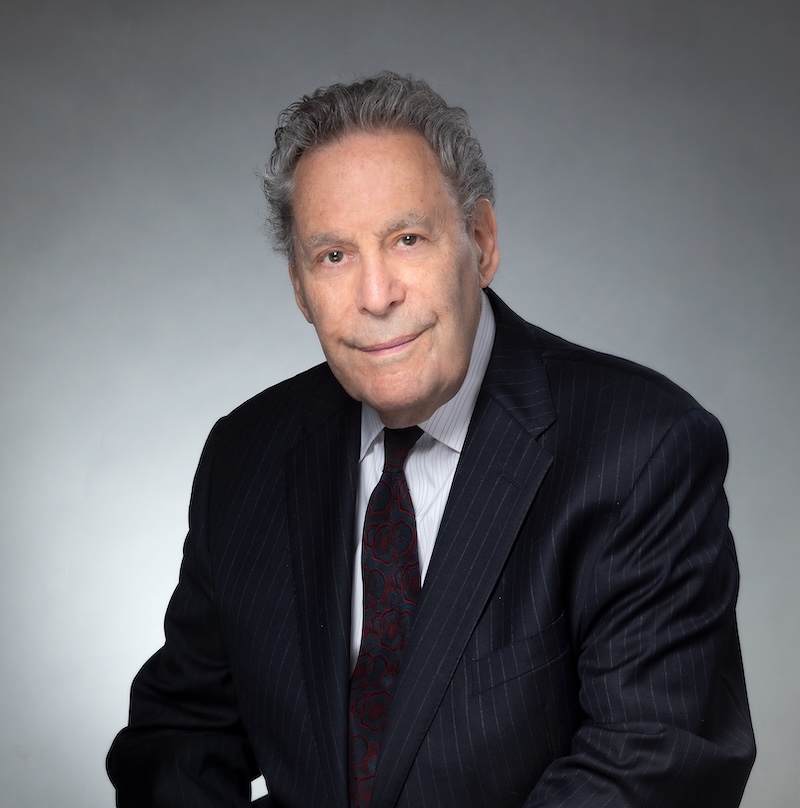
Doctor Smith graduated Temple University School of Dentistry in 1969 and completed a two-year tour of active duty as a captain in the U.S. Army Dental Corp. After practicing conventional dentistry for four years, Dr. Smith completed a two-year post-graduate orthodontic program in 1976. Sir Doctor Smith is a Knight Hospitaller, a dedicated professional organization dating back to the year 1050. The Knights Hospitallers have official recognition from the United Nations and the Pope for their tremendous humanitarian work with the poor. Doctor Smith is certified by the World Organization for Natural Medicine to practice natural medicine.
His broad base of post-graduate training has enabled him to integrate many healthcare specialties. He has accumulated an impressive list of credentials, which includes lecturing at Walter Reed Army Medical Center, the National Academy of General Dentistry, the Academy of Head, Neck and Facial Pain, Yonsei Memorial Hospital in Seoul, Korea, and dozens of guest lecture appearances at national and international symposia. He holds memberships and affiliations with a number of professional associations, including the International Associations for Orthodontists and the Academy of Head, Neck and Facial Pain. He has been an active member of the Holistic Dental Association since 1993, past president of the Holistic Dental Association, and editor of their professional journal from 2003 to 2006. He also served as past president of the Pennsylvania Craniomandibular Society.
Dr. Smith is a recognized international authority and pioneer in craniomandibular somatic disorders with a focus on resolving chronic pain. Dr. Smith was the first researcher in the world to document cranial bone motion by means of his groundbreaking research and development of the Dental Orthogonal Radiographic Analysis System. He was also the first researcher in the world to discover how to resolve chronic pain by removing tension patterns within the human skull by means of the Occlusal Cranial Balancing Technique. He is author of two landmark textbooks for professionals, Cranial-Dental-Sacral Complex and Dental Orthogonal Radiographic Analysis. He has also written an important book for the lay person, Headaches Aren’t Forever, a newly published book on CD-ROM, Alternative Treatments for Conquering Chronic Pain, and a second book on Reversing Cancer: A Survivor’s Guide for Understanding the Nature of Cancer, restoring the immune system, psychological healing, destroying cancer, and regeneration. Doctor Smith’s forty-eight years of clinical research have helped to identify several of the major missing links for successfully treating dentally related medical issues and chronic pain.
In addition, Dr. Smith has published over thirty articles and contributed chapters to several professional books. He is the developer of the Physiologic Adaptive Range Concept, Occlusal Cranial Balancing Technique, Quantum Testing Technique, and a special dental x-ray analysis system for measuring cranial bone motion. He holds two U.S. patents: a unique precision attachment for dental fixed bridgework and a flash adaptor to facilitate taking intraoral photographs. Doctor Smith is also president of the International Center For Nutritional Research, Inc., and he still maintains a private practice in Bucks County Pennsylvania, where he focuses his integrative healing concepts on chronic pain patients.
The concepts put forth in this article were designed to make healthcare practitioners, concussion victims, and their parents aware of advanced technologies that are presently available to help alleviate the chronic symptoms resulting from the structural distortions created by the concussion trauma. It must be understood that any physical damage to brain cells must be dealt with from a nutritional approach plus other modalities like scalar waves, soft lasers, hyperbaric oxygen therapy, and other energy healing concepts. The field of quantum energy healing will be the next major frontier in medicine.

