Twenty years of facial pain due to trauma and perpetuated by dental bridgework
Dental Bridgework / Face Pain Connection
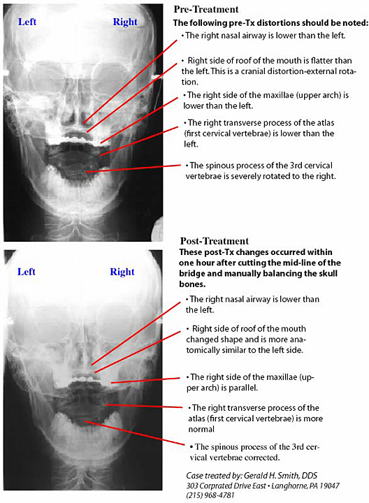
Pre and post-treatment x-ray views (below).
Case Report
Most people can sustain dental trauma and their body will recover to varying degrees. This miraculous ability to absorb physical force is designed into the human skull by means of a built in shock absorber system called sutures. These sutures are the connecting structures between the plates of the skull bones. There is soft tissue within these sutures comprised of fibers, blood vessels and nerves. In essence they are expansion/contraction joints that allows the head to compensate for physical trauma.
Miriam was in her twenties when she sustained an unfortunate accident. She was thrown from a horse drawn wagon and her face impacted on a roadside wooden fence. The end result was that five of her upper front teeth were fractured and required extraction. The physical impact also caused the plates of her skull to become distorted. Unfortunately this component of her traumatic episode was never addressed at the time of her emergency examination. This cranial distortion was also never treated until the patient came to my office twenty years later.
The emergency dental treatment involved restoring the upper missing teeth with fixed bridgework. The bridgework consisted of a cast gold framework with porcelain shaped like teeth and bonded to the gold. This prosthesis was then cemented on several of the remaining teeth, which were prepared to hold the fixed bridge. In reality this rigid device literally locked Miriam’s head into the distortion which occurred from the trauma. For twenty years this patient suffered headaches and facial pain from “unknown origin”.
Knowing how the craniosacral system works enables one to clearly understand the reason for the chronic pain – tension and sensory nerve stimulation within the dural membrane system inside the skull. The special x-ray system developed by this author helps to document the distortions present in the skull. The Dental Orthogonal Radiographic Analysis (DORA) was specifically developed to measure changes in the cranial structures before and after treatment. The before and after DORA x-rays of Miriam clearly depicts major changes that occurred following cutting the mid-line of the bridgework and adjusting the alignment of the cranial bones to remove the distortions created by the initial trauma. Cutting the mid-line of the existing bridgework was essential to allow freedom of the skull bones to move. Only then was it possible to realign and balance the skull. The end result was that the twenty-year facial and head pain resolved.

Knowing how the craniosacral system works enables one to clearly understand the reason for the chronic pain, tension and sensory nerve stimulation within the dural membrane system inside the skull.
FREE PRESENTATION
Download the slides from Dr. Smith's presentation on the Dental Whole Body Connection in Ontario on October 25, 2024.
A comprehensive seminar to awaken you to the potential dangers of dental treatments.

STAY INFORMED
Big tech and mainstream media try to suppress the powerful information I have to share. Subscribe here to stay informed!
